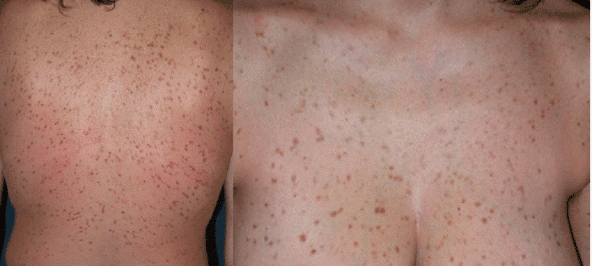
Mastocytosis is a rare condition characterized by the clonal proliferation and accumulation of altered mast cells (also known as tissue mast cells) in one or more organs. The most commonly affected sites include the skin, gastrointestinal tract, liver, spleen, and other internal organs.
Mastocytosis is divided into two main types:
-
Cutaneous mastocytosis (CM): affects only the skin
-
Systemic mastocytosis (SM): involves internal organs beyond the skin
Who Gets Cutaneous Mastocytosis?
CM is more common in children (about 65% of cases). In these cases, the disease is usually limited to the skin, has a benign course, and often resolves spontaneously by adolescence. Only a small percentage progresses to systemic disease.
In contrast, adults are more likely to be diagnosed with systemic mastocytosis, while isolated skin forms are rare (less than 5% of cases in adults).
There are three main types of cutaneous mastocytosis:
-
Maculopapular mastocytosis (including urticaria pigmentosa) – the most common
-
Diffuse cutaneous mastocytosis
-
Mastocytoma
Causes of Cutaneous Mastocytosis
Most cases are caused by gene mutations, which play a major role in both cutaneous and systemic forms of the disease. These mutations lead to an overproduction of mast cells, which release various bioactive mediators. This causes local or systemic inflammatory reactions.
Symptoms such as itching, redness, and swelling are typically caused by the release of these mediators.
Hereditary (familial) cases are extremely rare.
Symptoms and Clinical Presentation
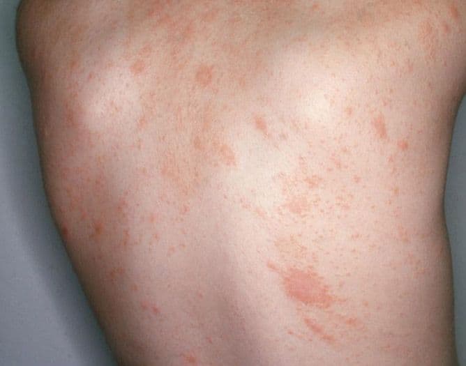
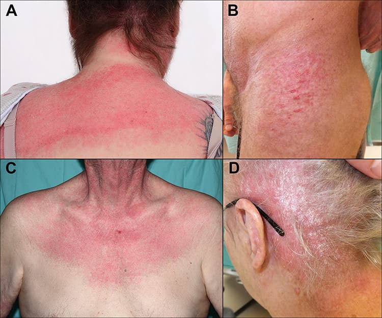
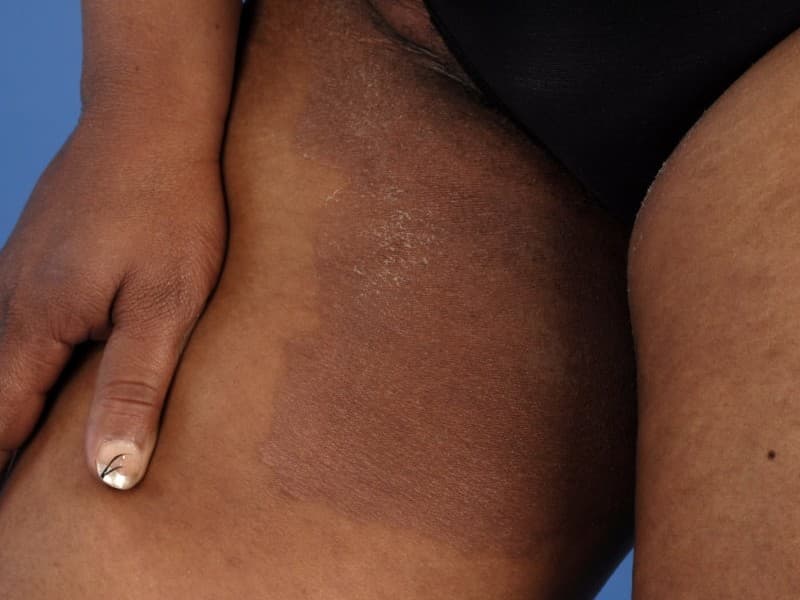
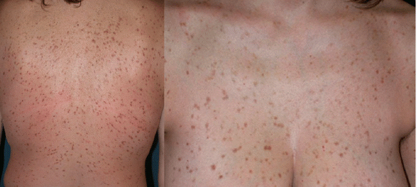
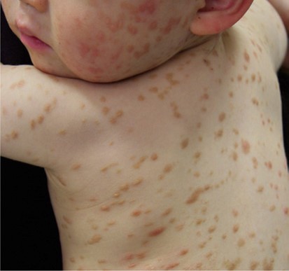
The most typical presentation is a gradually spreading rash with pink or brown macules and papules. Less commonly, plaques, nodules, blisters, or visible blood vessels (telangiectasias) may appear.
The most commonly affected areas include:
-
Chest, trunk, and limbs
-
The face, scalp, palms, and soles are usually spared
Common associated symptoms:
-
Itching
-
Flushing
-
Rapid heartbeat
-
Low blood pressure
-
Abdominal pain
-
Nausea or vomiting
Mastocytoma is a form of CM that usually occurs in children. It appears as a single, oval, reddish-yellow plaque or patch, typically on the chest, face, neck, or limbs.
A classic sign of CM is a positive Darier's sign: when the skin is gently rubbed, the lesion becomes red and swollen or may even form a small blister.
Diagnosis
Diagnosis is based on:
-
Medical history and clinical appearance
-
Dermoscopy to magnify and analyze lesion structure
-
In uncertain or widespread cases: skin biopsy for histological confirmation
In children, biopsy is rarely required due to the often self-limiting nature of the disease.
Additional tests may include:
-
Blood tests, including liver and kidney function
-
Serum tryptase level
-
Ultrasound of abdominal organs (to evaluate liver, spleen, and lymph nodes)
Treatment
In many cases, treatment focuses on symptom control and avoiding triggers rather than complete removal of lesions.
Main strategies include:
-
Educating the patient about triggers that may activate mast cells and how to avoid them
-
Carrying an emergency adrenaline autoinjector and knowing how to use it, as anaphylactic reactions can occur
-
Use of topical or systemic medications as needed
-
Phototherapy (e.g., UVB or PUVA) in selected cases
Common Triggers That Can Activate Mast Cells
Patients should avoid or be cautious with:
-
Insect stings (bees, wasps, jellyfish)
-
Physical friction or pressure (massage, rubbing, tight clothing)
-
Surgical procedures or biopsies
-
Certain medications (e.g., some anti-inflammatory drugs, contrast agents, some cardiac drugs)
-
Extreme temperatures – heat or cold
-
Stress, intense physical exertion, fever, or alcohol